How motion capture is revolutionising spinal care (Spinal Surgery News)

Kim Duffy, life sciences product manager at Vicon, looks at motion capture technology, which allows physicians to see how precisely patients enter their gait cycle, and gives an analysis of joint angles and movements, for when the spine is engaged, the lumbar, neck, thorax and head can all be affected.
Motion capture, the process of recording the movement of objects or people, was first introduced by the life sciences sector through its use for gait analysis in the early 1970s, and is now used widely by sports therapists, neuroscientists, and researchers for validation and control of computer vision and robotics.
Today, motion capture technology is at the frontline of cutting-edge clinical movement research, from helping stroke and amputee patients with rehabilitation, to understanding how to better treat those with cerebral palsy and arthritis. This technology is crucial to the work of leading research centres, universities, hospitals and private medical practices around the world.
One interesting area is the technology’s use in treating spinal-related issues, which is a common problem across the globe. According to both the American Chiropractic Association (ACA) [1] and World Health Organization (WHO) [2], back pain is the single leading cause of disability worldwide, with one in three people stating that back pain impacts their everyday life [3].
But through the use of motion capture to digitally track a person’s motions, physicians are able to analyse a wide range of back problems such as generative disc disease, chronic lower back and neck pain, and scoliosis in adults and children. Having the ability to diagnose these back-related issues is helping physicians all over the world to identify the best treatment approaches and help to improve not just patient care, but also quality of life for hundreds of thousands of patients.
Motion capture in a laboratory setting
The Texas Back Institute (TBI) in the USA is a private multi-disciplinary spine care centre dedicated to improving the lives of patients through research knowledge and wide-ranging treatment approaches, and is also a key example of how motion capture technology is being used to revolutionise spinal care.
In order for TBI’s team of physicians to address a wide range of spinal-related problems, they needed as much information as possible. Data lies at the heart of the optimal treatment decisions, whether they are surgical or nonsurgical in nature.
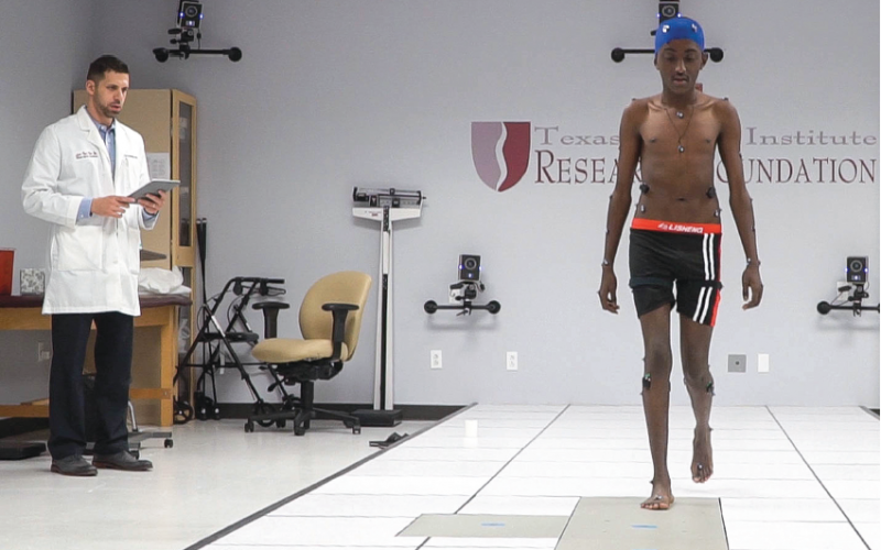
This led Dr Ram Haddas, TBI’s Director of Research to build an advanced motion capture laboratory setting using Vicon equipment that would allow them to capture information about a person’s spine and produce research that had a major impact on the analysis and treatment of commonly performed spine surgeries carried out at the centre. Dr Haddas has evaluated more than 600 patients and used this work to drive much published research on this topic.
With the use of motion capture cameras and video cameras in a lab setting, physicians are able measure a patient’s gait and balance, as well as assess the pain scale.
Pain, what patients feel and how it’s measured, is a critical part of the assessment and treatment process, and is often measured by a highly subjective rating system where patients rate discomfort on a one-to-ten scale. While every patient has a different perception of pain, combining data from both the motion capture system and electromyography (EMG) can help to quantify how pain affects mobility and establish more objective criteria. Additionally, by utilising motion capture, Dr. Haddas, was the first to quantify the dimensions and compensation strategy of the Cone of Economy (efficiency level in a quiet stand).
Physicians can also compare, for example, what a patient reports with how fast they’re walking or their range of motion. There is a proven relationship between physical and mental states, and through data and analysis, medical teams are able to scientifically correlate the two together. Motion capture can then be used to prove the relationship between the patient’s pain and their movement.
Medicine in motion
At TBI, candidates for surgical procedures generally undergo testing one week before their operation for a baseline functional evaluation, and then return for a short-term follow up before finishing with long-term visits at one and two years post-surgery.
Rather than focus on traditional treadmill analysis, patients walk at their comfortable speed in the lab that mimics real-world movement conditions. While surgery is traditionally based on static imaging, as soon as patients start to move, things change. The motion capture system enables physicians to see precisely how patients enter their gait cycle, and even sway while standing, and enables analysis of joint angles and movements. When the spine is engaged, the lumbar, neck, throat and head can all be affected.
Using a full-body marker set and the motion capture system, the surgical team is able to collect data on gait, walking speed, cadence, balance and joint angles – including the ankle, knee and hip.
EMG data measuring electrical activity produced by the muscles is also fully integrated within the motion capture system to provide physicians with a full image of how much muscle energy a patient is expending in the gait cycle.
With height, weight and other measurements, including lifting and balance details, the physicians can also calculate a patient’s exact centre of mass and displacement. During a one-minute test, physicians can track the extent of displacement of the centre of mass of a scoliosis patient for example, which averages almost a full metre, while a control subject sways significantly less. These are all made possible for the first time through the centre’s motion capture lab setting.
Once testing is complete, all data is processed using an auto-labelling technique, and detailed reports are then generated for physicians, and also used for research into treatment decisions.
Inspiring outcomes
These reports generated by the lab significantly help with patients’ diagnoses and establishing controls, pre and post-surgery, for both the short and long term. For surgical patients, one-year reports can be compared not only to pre-surgery reports, but also to healthy control subjects.
Vicon is continuing to push the boundaries of technology, and by working with spinal care centres around the globe, is helping to further deepen our understanding of human motion to better diagnose, treat, rehabilitate and track spinal conditions – significantly improving the lives of many.
References:
https://www.acatoday.org/Patients/What-is-Chiropractic/Back-Pain-Facts-and-Statistics

 Tiger Buford – retained recruiter dissecting orthopedics